Q. The talus bone of the foot gets the load of the body from the tibia. The talus bone then distributes this weight towards the ground in two Instructions: 1-50 percent of the body excess weight is passed in a very posterior course and a person-fifty percent of the load is passed in an anterior route.
The interosseous border of every bone could be the attachment web page for your interosseous membrane in the leg, the connective tissue sheet that unites the tibia and fibula.
The distal close of the femur has medial and lateral bony expansions. On the lateral side, The sleek part that covers the distal and posterior aspects of the lateral enlargement is the lateral condyle of the femur. The roughened area on the outer, lateral facet of your condyle could be the lateral epicondyle with the femur. Equally, The sleek region from the distal and posterior medial femur would be the medial condyle in the femur, as well as the irregular outer, medial side of this is the medial epicondyle of your femur. The lateral and medial condyles articulate While using the tibia to variety the knee joint.
The hardware attaches directly to existing new style quickie frames, and buyers can decide on the pad size and aspect of support.
The two heads of your four dorsal interossei arise on two adjacent metatarsals and merge within the middleman Areas. Their distal attachment is within the bases from the proximal phalanges of the 2nd-fourth digits. The interossei are structured with the next digit as being a longitudinal axis; the plantars work as adductors and pull digits three–5 to the next digit; whilst the dorsals work as abductors. Moreover, the interossei work as plantar flexors on the metatarsophalangeal joints. Last of all, the flexor digitorum brevis arises from beneath the calcaneus to insert its tendons on the center phalanges of digit two–four. Because the tendons with the flexor digitorum longus run between these tendons, the brevis is typically referred to as perforatus. The tendons website of both of these muscles are surrounded by a tendinous sheath. The brevis functions to plantar flex the middle phalanges.[36]
The quadriceps will be the knee extensor, though the rectus femoris Moreover flexes the hip joint, and articular muscle with the knee safeguards the articular capsule of your knee joint from being nipped all through extension. The sartorius runs superficially and obliquely down to the anterior facet of the thigh, from your anterior excellent iliac spine on the pes anserinus within the medial side of the knee, from in which it truly is even further prolonged into your crural fascia. The sartorius functions as a flexor on each the hip and knee, but, as a result of its oblique system, also contributes to medial rotation on the leg as among the list of pes anserinus muscles (While using the knee flexed), also to lateral rotation with the hip joint.[23]
Patellofemoral syndrome may be initiated by many different will cause, including person versions in The form and motion of your patella, a direct blow on the patella, or flat toes or incorrect footwear that induce excessive turning in or out on the toes or leg.
Stretching ahead of demanding Bodily action has become imagined to raise muscular effectiveness by extending the comfortable tissue previous its attainable duration if you want to increase range of movement.
If stretching with the ligaments is extended, excessive, or repeated, it may lead to a gradual lengthening from the supporting ligaments, with subsequent depression or collapse with the longitudinal arches, particularly to the medial side on the foot. This ailment lower limb supports is known as pes planus (“flat foot” or “fallen arches”).
The talus bone in the foot gets the weight of the human body from your tibia. The talus bone then distributes this body weight toward the bottom in two directions: a single-half of your body bodyweight is handed inside a posterior course and 1-50 % of the burden is handed in an anterior way.
Veins of your leg The veins are subdivided into three systems. The deep veins return around eighty five p.c of the blood as well as the superficial veins somewhere around fifteen p.c. A series of perforator veins interconnect the superficial and deep techniques.
roughened space over the posterior aspect in the proximal femur, extending inferiorly in the foundation with the increased trochanter
Nearly all the thigh muscles, the "accurate" thigh muscles, insert around the leg (either the tibia or the fibula) and act totally on the knee joint. Frequently, the extensors lie on anterior with lower limb supports the thigh and flexors lie to the posterior. While the sartorius flexes the knee, it can be ontogenetically regarded an extensor considering that its displacement is secondary.[15]
Buyers of our products and solutions ought to feel confident, secure, and built-in with their prosthetic leg, knee, ankle, foot or limb system; in a position to Reside their existence because they picture. The individual’s lower limb performs just like a normal extension on the human human body.
Celebrity Then and Now
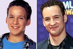 Ben Savage Then & Now!
 Marla Sokoloff Then & Now!
 Daryl Hannah Then & Now!
 Heather Locklear Then & Now!
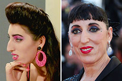 Rossy de Palma Then & Now!